Imaging techniques
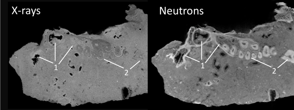
I contributed to the analysis of one of the first fossil primates scanned by neutron radiation that provides enhanced contrast (see link).
In 2019, we scanned the skull of ‘Little Foot’ at the Diamond Light Source synchrotron (UK) and published the first paleohistological observations of a hominin specimen older than 3 million years (see link).

Analytical tools
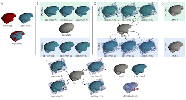
I contributed to the development of the surface deformation method, which eliminates the need for landmark points traditionally used in geometric morphometrics. In addition to the endocasts (see link), I applied this method to crania (see link) as well as vertebrae (see link).

Because of the subjective nature of identifying brain imprints in endocasts, we developped a tool for the automated detection of sulcal imprints (see link) that is available here!

Using existing methods developped in neuroscience, we published and shared (here) the first virtual atlas that maps the variation of brain sulci in human endocasts (see link).
